APPLICATION
SRS射頻信號發生器在金剛石氮空位色心的研究介紹
量子計算和量子傳感近年來受到了廣泛的關注.金剛石氮空位色心以其簡單穩定的自旋能級結構、高效便捷的光學躍遷規則以及室溫下超長的自旋量子態相干時間而成為量子信息科學中引人矚目的新星,近十年來,金剛石氮空位色心的研究呈爆炸式增長(見圖1)
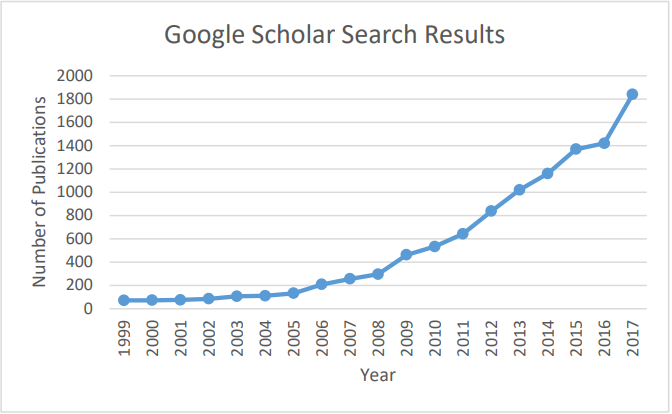
圖1:每年提及“氮空位”和“金剛石”的出版物總數(Google Scholar)。
顧名思義,氮空位色心(NV-Nitrogen-Vacancy center)(圖2)是由一個氮原子取代金剛石中的一個碳原子,然后捕獲周圍的一個空穴形成的。在NV位上懸浮鍵的電子通過雜化產生自旋-1系統,具有明確的能量狀態和較長的自旋壽命,即使在室溫下也是如此。這樣就可以通過磁共振的自旋操控技術在精確定時的微波磁場下來控制NV點的自旋狀態。由于它的能級對外部磁場和電場都很敏感,使得這種自旋狀態可以作為外部環境的傳感器。
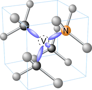
圖2:金剛石中NV色心由碳原子(灰色)、氮原子(橙色)和相鄰空位組成,并有懸空鍵(紫色)[1]。
在NV實驗中監測到的能量狀態如圖3所示:
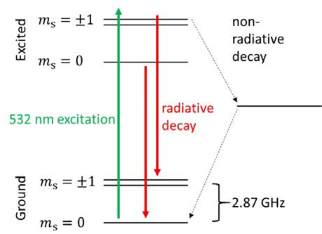
眾所周知,金剛石中的缺陷會使原本透明的晶體產生顏色(例如,氮取代缺陷使金剛石呈現黃色,而硼取代則產生藍色)。在532nm激光激發下,NV在637 nm處產生熒光衰減。重要的是?s=±1自旋態是無輻射衰減,從而提供了一種利用監測光致發光變化來檢測自旋狀態的方法。也就說,當自旋態被激發到不穩定態?s =±1級(使用2.87 GHz微波),有缺陷的紅光輸出會略有減少(圖4 (a))。此外,?s =±1的能級狀態可以被外部磁場調節。通過測量吸收峰之間的頻率分裂(如圖4(b)所示),可以測量出該外部磁場。
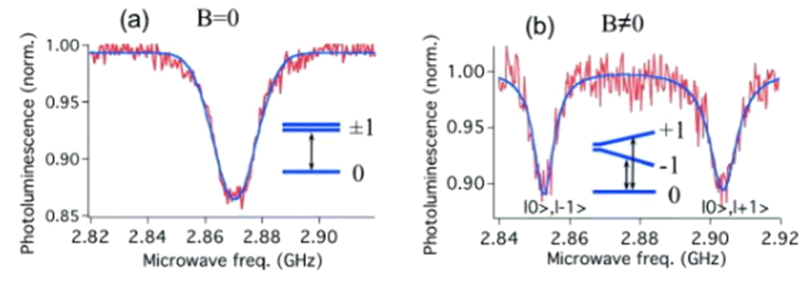
圖4。(a)在2.87GHz躍遷共振微波作用下的光致發光衰減(??= 0和1狀態之間)。
(b)非零磁場下的??= +1和-1狀態之間的分裂[3]。
能量狀態的改變也可能是由外部電場[4],溫度[5] [6]或金剛石晶格的應變[7]引起,這使得NV光學探測磁共振(ODMR)對于感測這些量中的任何一個都有作用。因為NV軸可以匹配金剛石晶格中4個晶軸中的任何一個,因此可以執行3D矢量場感測[8]。例如,結合共聚焦顯微鏡技術可以解決NV共振位移的空間分辨圖,趨磁細菌產生的磁場矢量圖譜詳見(圖5)[g]。
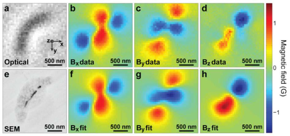
圖5。(a)趨磁細菌的光學圖像。(e)同一細菌的SEM圖像,其中磁性納米粒子(黑色)清晰可見。(b,c,d)磁場x, y, z分量由NV金剛石光學探測磁共振測量確定。(f,g,h)實測場分布的數值擬合(來自[g])。
和大多數磁共振測量一樣,第一次NV實驗涉及大量的缺陷。然而,令人驚訝的是,在低缺陷密度的樣品中,單個NV色心可以被隔離和監測。由于NV是原子大小的,這意味著單自旋NV傳感器的空間分辨率僅受限于與其感興趣樣品的距離。例如,通過在原子力顯微鏡探針的尖端定位單個NV色心,已經證明了具有25nm分辨率和56nT/Hz-1/2的磁疇成像[10]。
此外,NV色心的極端敏感性和生物相容性(金剛石只是碳,因此沒有表現出毒性),顯示了納米核磁共振測量的特殊前景,其中NV中心被耦合到與之感興趣的有機和生物分子的核自旋的雜散場上[11]。因此,NV被定位為革命性的納米磁共振成像技術,并提供了一種在環境條件下獲得單分子3D圖像的途徑[12]。事實上,NV色心的單質子自旋探測已經被證實[13]。
NV場感測和納米成像不限于靜態場。現在,一些研究小組已經證明了基于NV探測和時變磁場的磁場掃描是由鐵磁鐵中的自旋波產生[14][15]或電流波動[16][17]。
為了進一步增加NV應用的長列表、長壽命的自旋狀態、與外部場的可操作性以及與環境的可調諧相互作用(通過晶格工程)也使它們成為量子計算中信息單元“qubit”的領跑者[18]
量子計算和傳統計算一樣,可能需要將長壽命存儲位與快速處理位分離。將長壽命的“內存”量子位(基于附近的氮原子或13c原子核自旋)與NV自旋的耦合[19][20][21]可以精確地提供這種內存和處理架構。超導磁通量子位(另一個強大的量子計算候選者)和NV色心之間的相干耦合也得到了證明[22],為量子處理和存儲所需要的光學與微波之間讀寫的接口鋪平了道路。
SRS產品在NV研究中的應用
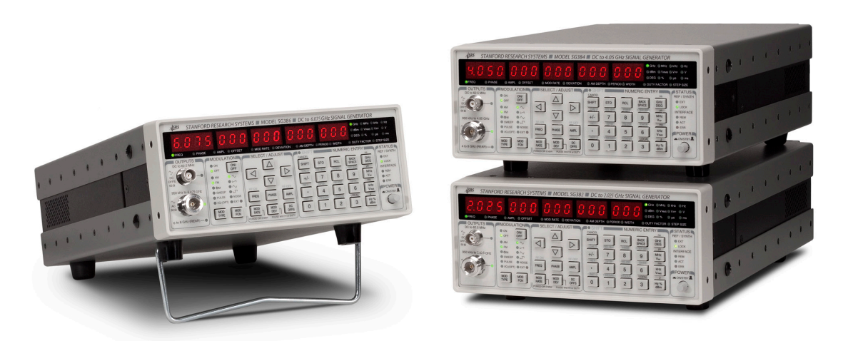
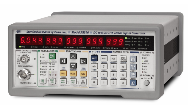
在NV金剛石中進行光學檢測磁共振實驗的關鍵是掃描微波頻率的能力。為了測量自旋態壽命,執行微波脈沖序列也很有用。SRS射頻信號發生器(SG384和SG386)和矢量信號發生器(SG394和SG396)非常適合在NV-ODMR研究中的所需要的頻率范圍內產生微波。下面是使用SRS產品的NV相關參考的列表。
1. Optical magnetic imaging of living cells
https://www.nature.com/articles/nature12072
使用設備: SG384 (+ amplifier)
2. Miniature Cavity-Enhanced Diamond Magnetometer
https://journals.aps.org/prapplied/abstract/10.1103/PhysRevApplied.8.044019
使用設備: SG394 (+ 16W amplifier, circulator)
3. Microwave-free magnetometry with nitrogen-vacancy centers in diamond
https://aip.scitation.org/doi/full/10.1063/1.4960171
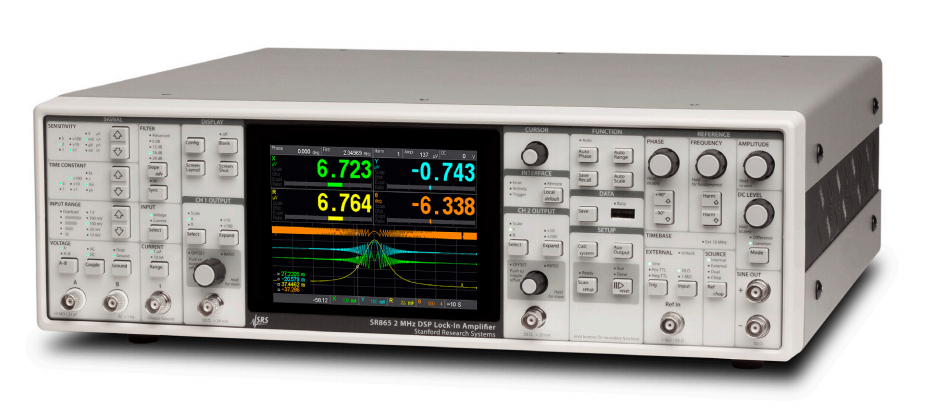
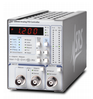
使用設備: SR865, SIM960
 |
 |
4. Precision temperature sensing in the presence of magnetic field noise and vice-versa using
nitrogen-vacancy centers in diamond
https://arxiv.org/abs/1802.07224
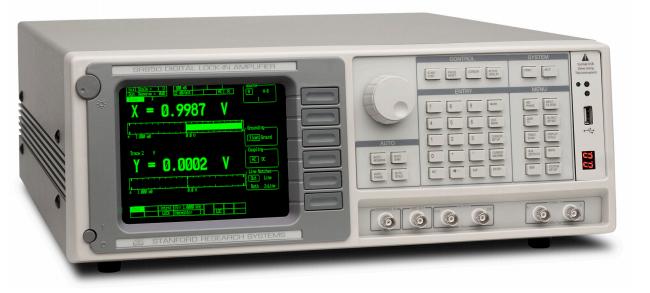
使用設備: SG394(2x) (+power combiner, switch, and amplifier), SR850
5. Quantum Control of Nuclear Spins Coupled to Nitrogen-Vacancy Centers in Diamond
(dissertation, S. Sangtawesin)
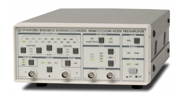
使用設備: SG394 (+ amplifier)
 |
 |
References
[1] Z. Fotoniki, "Diamonds with nitrogen-vacancy (NV) color centres," 2018. [Online]. Available:
https://zf.if.uj.edu.pl/en/node/518.
[2] R. M. Teeling-Smith, Single Molecule Electron Paramagnetic Resonance and Other Sensing
And Imaging Applications with Nitrogen-Vacancy Nanodiamond, Columbus, Ohio: OhioLink,
2015.
[3] J.-F. Roch, "Optical determination and magnetic manipulation of a single nitrogen-
vacancy color center in diamond nanocrystal," Advances in Natural Sciences: Nanoscience
and Nanotechnology, 2010.
[4] J. Wrachtrup, "Electric-field sensing using single diamond spins," Nature Physics, vol. 7, pp.
459-463, 2011.
[5] J. Wrachtrup, "High-Precision Nanoscale Temperature Sensing Using Single Defects in
Diamond," Nano Letters, vol. 13, pp. 2738-2742, 2013.
[6] M. D. Lukin, "Nanometre-scale thermometry in a living cell," Nature, vol. 500, pp. 54-58,
2013.
[7] S. Prawer, "Electronic Properties and Metrology Applications of the Diamond NV- Center
Under Pressure," Physical Review Letters, vol. 112, p. 047601, 2014.
[8] D. D. Awschalom, "Vector magnetic field microscopy using nitrogen vacancy centers in
diamond," Applied Physics Letters, vol. 96, p. 092504, 2010.
[9] R. L. Walsworth, "Optical magnetic imaging of living cells," Nature, vol. 496, pp. 486-489,
2013.
[10] A. Yacoby, "A robust scanning diamond sensor for nanoscale," Nature Nanotechnology,
2012.
[11] D. Rugar, "Nanoscale Nuclear Magnetic Resonance with a Nitrogen-Vacancy Spin
Sensor," Science, vol. 339, pp. 557-560, 2013.
[12] S. Castelletto, "Towards Single Biomolecule Imaging via Optical Nanoscale Magnetic
Resonance Imaging," Small, vol. 9, p. 4229, 2015.
[13] A. O. Sushkov, "Magnetic Resonance Detection of Individual Proton Spins Using Quantum
Reporters," Physical Review Letters, vol. 113, p. 197601, 2014.
[14] C. S. Wolfe, "Spatially resolved detection of complex ferromagnetic dynamics using
Optically detected nitrogen-vacancy spins," Applied Physics Letters, vol. 108, p. 232409, 2016.
[15] C. Du, "Control and local measurement of the spin chemical potential in a magnetic
insulator," Science, vol. 357, pp. 195-198, 2017.
[16] S. Kolkowitz, "Quantum electronics. Probing Johnson noise and ballistic transport in
normal metals with a single-spin qubit," Science, vol. 347, pp. 1129-1132, 2015.
[17] A. Waxman, " Diamond magnetometry of superconducting thin films," Physical Review B,
vol. 89, p.054509, 2014.
[18] R. H. a. L. Childress, "Diamond NV centers for quantum," MRS Bulletin, vol. 38, pp. 124-
138, 2013.
[19] M. D. Lukin, "Quantum Register Based on Individual Electronic and Nuclear Spin Qubits
In Diamond," Science, vol. 316, pp. 1312-1316, 2007.
[20] D. D. Awschalom, "A quantum memory intrinsic to single nitrogen–vacancy centres in
diamond," Nature Physics, vol. 7, pp. 789-793, 2011.
[21] M. D. Lukin, "Room-Temperature Quantum Bit Memory Exceeding One Second," Science,
vol. 336, pp. 1283-1286, 2012.
[22] K. Semba, "Coherent coupling of a superconducting flux qubit to an electron spin
ensemble in diamond," Nature, vol. 478, pp. 221-224, 2011.
[23] C. Degen, "Single-proton spin detection by diamond magnetometry," Science, 2014.
Copyright ? 2020 Zolix .All Rights Reserved 地址:北京市通州區中關村科技園區通州園金橋科技產業基地環科中路16號68號樓B.
ICP備案號:京ICP備05015148號-1
公安備案號:京公網安備11011202003795號

 13810146393
 在線咨詢
在線咨詢